Gerald Obermair Professorship of Physiology at KL University
At the beginning of March, the Department of Physiology started at the KL with the Professor of Physiology, Dr. Gerald Obermair.

Gerald Obermair (born 1973 in Salzburg) studied Zoology and Neuroscience at the University of Salzburg and the Bowling Green State University (Ohio). After his dissertation in the field of cellular neurobiology at the Universities of Innsbruck and Salzburg he worked at the Medical University of Innsbruck at the Section of Physiology (Habilitation 2010), since 2013 as Associate Professor. Together with other scientists, Obermair is project leader in the Collaborative Research Centre "Cell signaling in chronic CNS disorders".
In 2018 he was able to recruit the doc.funds PhD program "CavX - Calcium channels in excitable cells" as spokesperson.
https://www.uibk.ac.at/pharmazie/pharmakologie/sfb-f44/ https://www.i-med.ac.at/mypoint/news/714221.html https://www.i-med.ac.at/mypoint/news/720543.html
Prices:Sackler Research Award of the University of Salzburg 1998Sanofi-Aventis Prize 2005
Scientific societies:Austrian Neuroscience Association (Board member from 2012-2016)ANA representative within the FENS governing council (since 2012)Society for Neuroscience
Expert activity:Journals (selection): Biophysical Journal, Cellular and Molecular Life Sciences, Cerebral Cortex, European Journal of Neuroscience, Journal of Neurochemistry, The Journal of Neuroscience, Nature communications, PNAS, Scientific ReportsFunding organizations (selection): ANR (France), APVV (Slovakia), Parkinson's UK
Journal cover selections:EJN 2004, JBC 2010, JN 2010, JN 2014, JN 2019https://www.i-med.ac.at/dpmp/physiologie/research/obermair/gallery.htmlhttp://www.jneurosci.org/content/39/14.cover-expansionhttp://www.jneurosci.org/content/39/14/2573 Main research areas:Investigation of the manifold functions of neuronal calcium channels in muscle and nerve cells. In particular, Obermair investigates the role of the individual regulatory calcium channel components in the formation and stability of synapses in healthy and diseased (e.g. Parkinson's or autism) nerve cells.
Info on research areashttps://www.i-med.ac.at/mypoint/news/686749.htmlhttps://www.i-med.ac.at/pr/docs/medinn0117.pdf (title page and articles)
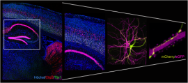
Zooming into the mouse brain: From cytoarchitecture to synaptic connections. The two pictures on the left show adult mouse brain sections which were analyzed via labelling of Ctip2 (red), Tbr1 (green) and nuclei (blue). The overview picture on the right illustrates how two hippocampal neurons, transfected with mCherry or eGFP (depicted in yellow and magenta), interact with each other. The detail further demonstrates how a mCherry positive presynaptic nerve terminal forms synaptic contacts along the postsynaptic dendrite of an eGFP positive neighbor neuron.


