News
11.12.2023
New scientific publication - Simultaneous Dry and Gel-Based High-Density Electroencephalography Recordings
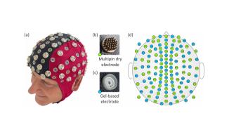
EEG caps with high-density dry sensors have only been evaluated in sequential measurements, but not in simultaneous measurements. For a first comparison of the performance of gel and dry electrodes in simultaneous EEG measurements, we designed and realized a new EEG cap consisting of 64 gel and 64 dry electrodes. In a pilot study with ten subjects, we recorded and analyzed electrode-skin impedances, resting EEG, triggered eye blinks, and visual evoked potentials (VEPs). To overcome the problem of different electrode positions when comparing simultaneous measurements, we performed a spatial frequency analysis of the simultaneously recorded EEGs using Spatial Harmonics Analysis (SPHARA).
The project leading to this publication was carried out in cooperation between scientists from the Department of Biostatistics and Data Science at KL (U. Graichen) and the Institute of Biomedical Engineering and Computer Science at TU Ilmenau (P. Fiedler, E. Zimmer, J. Haueisen).
Fiedler P, Graichen U, Zimmer E, Haueisen J. Simultaneous Dry and Gel-Based High-Density Electroencephalography Recordings. Sensors. 2023; 23(24):9745. https://doi.org/10.3390/s23249745
30.10.2023
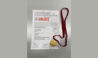
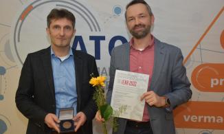
Certificate of Honor and Gold Medal for a New Patent in the Field of Neuroscience at the iNEA International Exhibition in Nuremberg
On October 30th, a patent was awarded with a certificate of honor and a gold medal at the international trade fair iNEA in Nuremberg. The patent is the result of a collaboration between scientists from the Department of Biostatistics and Data Science at KL (U. Graichen) and the Institute of Biomedical Engineering and Informatics at TU Ilmenau (A. Hunold, J. Haueisen).
The award-winning invention can be used for non-invasive transcranial electrical stimulation of target areas in the brain and is of interest for the treatment of depression and neurorehabilitation after stroke. The patented approach makes it possible to optimally distribute the stimulation current to the electrodes and thus address a specific target area in the brain.
13.06.2023
New scientific publication - Spatiotemporal phase slip patterns for visual evoked potentials, covert object naming tasks, and insight moments extracted from 256 channel EEG recordings
The aim of our research project was to investigate how phase transitions of coordinated activity of cortical neurons behave during visual object naming tasks. In conventional EEG analyses, such as power spectral density (PSD) calculations, the small effects of phase transitions remain hidden in the PSD of the bulk of the EEG. In this research project, we have developed and implemented a data processing chain to extract these phase transitions. The results of our research project show that phase shifts and phase shift rates (PSRs) complement the EEG results. Phase jump activity is often observed in different areas compared to the spatial patterns of the EEG. This suggests that a combined analysis with spatial representations of EEG and PSRs provides a more comprehensive picture of the underlying brain activity.
Ramon C, Graichen U, Gargiulo P, Zanow F, Knösche TR and Haueisen J (2023) Spatiotemporal phase slip patterns for visual evoked potentials, covert object naming tasks, and insight moments extracted from 256 channel EEG recordings. Front. Integr. Neurosci. 17:1087976. doi: 10.3389/fnint.2023.1087976
Quantitative analysis of risk-taking using online gaming
Overstimulation by dopamine can lead to impulse control disorders in patients and to increased risk-taking and hasty decisions. These symptoms can also affect patients with Parkinson's disease who are treated with certain drugs (e.g., dopamine agonists).
In a joint project of the two scientists OÄ Dr Stephanie Hirschbichler, MSc PhD from the University Hospital St. Pölten, Department of Neurology and Dr Uwe Graichen from the Department of Biostatistics and Data Science, an online game was implemented that enables a quantitative analysis of the risk tolerance of affected patients.
To the news article
24.01.2023
Post in the biostatistics blog on survival analysis
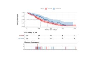
We have published a new post on the topic of survival analysis in our biostatistics blog. Survival analysis involves statistical procedures that compare the time to a specific event between groups to estimate the effect of prognostic factors such as medical treatment or harmful influences. The event may be the death of the patient. It can also be any other endpoint, such as recovery or the occurrence of a complication. In this post, we present three common survival time analysis methods and how they are performed using Gnu R: the Kaplan-Meier survival time curve, the log-rank test and Cox regression. The blog post can be found here.
07.12.2022
Granting and award of a new patent in the field of neurosciences
In the field of neurosciences, especially for the objective determination of individually perceived scattered light events, a team of inventors from the Technical University of Ilmenau (B. Solf, S. Schramm, D. Link, S. Klee) under the lead of Prof. Klee succeeded in filing a successful application and obtaining a process and device patent: "Method and device for the objective electrophysiological determination of the perceived scattered light of the human eye". At the same time, we were able to present the patent at this year's international inventors' trade fair iENA in Nürnberg and have it evaluated by a jury of experts. The award of a bronze medal was received on the 29th of November. The know-how on which the patent is based is to be further developed in the future, for example in an ongoing cooperation project with the Technical University of Ilmenau.
23.09.2022
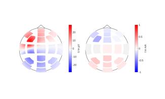
Disclosure of the patent "Verfahren und System zur Erzeugung und Applikation von Stimulationskonfigurationen am Menschen sowie dazugehöriges Computerprogramm "
On September 22nd 2022, the patent "Method and system for the generation and application of stimulation configurations in humans and associated computer program" was disclosed by the German Patent and Trade Mark Office. The patented invention was developed as part of a cooperation between scientists from the Department of Biostatistics and Data Science at KL and the Institute of Biomedical Engineering and Computer Science at TU Ilmenau. It can be used for non-invasive transcranial current stimulation of target areas in the brain and is of interest for the therapy of depression and neurorehabilitation after stroke, among other things. With the help of the newly developed approach, it is possible to distribute the stimulation current optimally to the electrodes and thereby address a dedicated target area in the brain. In the invention, a spatially harmonic analysis of the current topographies is used to calculate the optimal current distribution for multi-channel transcranial current stimulation and to determine a suitable electrode configuration.
22.09.2022
New associate researcher joins the team of the Department of Biostatistics and Data Science
Lydia Hofmann, M.Sc. supports the Department of Biostatistics and Data Science of the KL as an associate researcher and is working on a project with the aim of improving source localization using EEG
Starting in July, Ms. Lydia Hofmann, M.Sc., with her expertise in the fields of medical measurement technology and data analysis, joined the team of the Department of Biostatistics and Data Science at KL as an associate researcher. She will be conducting research in the field of source localization using electroencephalography as part of a project funded by the German Research Foundation. Ms. Hofmann has an additional affiliation at the Department of Optoelectrophysiological Medical Engineering of the Institute of Biomedical Engineering and Computer Science at TU Ilmenau. With her employment, a research cooperation agreement signed between KL and TU Ilmenau in March 2022 will be brought to life.

20.07.2022
Biostatistics Blog
The Department of Biostatistics and Data Science of the KL now runs a statistics blog. In the blog, frequently occurring questions arising in the fields of biostatistics and data science are addressed. Based on a practical example, a step-by-step solution to the specific question is presented, which can serve the reader of the blog as a blueprint for his or her own analyses. Gnu R is usually used for the exemplary solution of the biostatistical questions. In the individual blog entries, in addition to the practical solution, the theoretical basis of the presented problem is briefly discussed, and the used Gnu R toolboxes are introduced. The KL Statistics Blog can be accessed here.
14.03.2022
Cooperation agreement between KL and TU Ilmenau
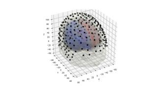
Joint cooperation project between the Department of Biostatistics and Data Science of the KL and the Institute of Biomedical Engineering and Computer Science of the TU Ilmenau with the aim to develop a new source localization method in EEG
On March 14th, 2022, a research cooperation agreement was signed between the Departments of Biostatistics and Data Science of KL and the Institute of Biomedical Engineering and Computer Science of TU Ilmenau. The goal of the first joint research project of the two cooperation partners is the development of an efficient and robust localization method for sources in the brain based on EEG data. The project is led by Dr. Uwe Graichen (KL) and worked on by a PhD student of the TU Ilmenau. Through the newly established cooperation, the expertise in the fields of bioelectromagnetism and model-based analysis of the TU Ilmenau and in the fields of biostatistics and data analysis of KL can be beneficially combined for both sides.
02.02.2022
New release of the Python toolbox SpharaPy published
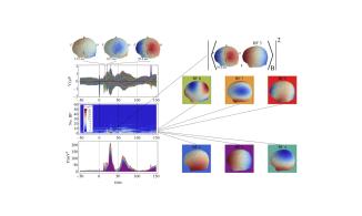
A new release of the Python toolbox SpharaPy has been published by members of the division of Biostatistics and Data Science of the Karl Landsteiner University of Health Sciences. SpharaPy is a Python implementation of the new approach for spatial harmonic analysis (SPHARA) that extends the classical spatial Fourier analysis to non-uniformly positioned samples on an arbitrary surface. The SpharaPy toolbox provides classes and functions to determine the SPHARA basis functions, to perform data analysis and synthesis (SPHARA transform) as well as classes to design spatial filters using the SPHARA basis. In the field of medical data science, SpharaPy is mainly used for spatial analysis of EEG data. In the current release, smaller bug fixes have been incorporated, the efficiency of the toolbox has been improved and adjustments have been made to new versions of the toolboxes used by SpharaPy.
https://www.sciencedirect.com/science/article/pii/S2352711019301670
https://gitlab.com/uwegra/spharapy
https://spharapy.readthedocs.io/en/latest/
https://pypi.org/project/SpharaPy/
23.12.2021
New publication on data-driven, three-dimensional light field imaging of the human retina and depth measurement of the optic disc.
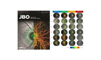
In the publication "3D retinal imaging and measurement using light field technology", together with a team of researchers from Germany, we have established for the first-time three-dimensional imaging of the ocular fundus without the use of laser technologies. Light-field fundus photography has the potential to be a new milestone in ophthalmology. Up-to-date publications show only unsatisfactory image quality, preventing the use of depth measurements. We show that good image quality and, consequently, reliable depth measurements are possible, and we investigate the current challenges of this novel technology. The measurement of the depth of the optic nerve head is in good agreement with both model measurements and established optical coherence tomography (OCT). Thus, the new technology can be a contribution to early diagnosis of e.g. glaucoma.
Stefan Schramm, Alexander Dietzel, Dietmar Link, Maren-Christina Blum, Sascha Klee, "3D retinal imaging and measurement using light field technology," J. Biomed. Opt. 26(12), 126002 (2021), doi: 10.1117/1.JBO.26.12.126002.
http://dx.doi.org/10.1117/1.JBO.26.12.126002.
Figure: Light field images of all subjects and wavelengths. Total focus images with the corresponding depth map overlays and measurement points normalized to the maximum per subject are shown. Most measurement points were found at a wavelength of 520 nm (for subjects S1 to S3, S5, and S6). Subject S4 had slightly more found measurement points at 600 nm. The internal fixation needle in the first intermediate image plane of the fundus camera (straight line within the fundus images) is imaged at S1 to S4 and S6 and depth-estimated thereon.
30.09.2021
Conference paper and talk – European Association for Vision and Eye Research (EVER2021)
“Neuronal sources of visually evoked potentials using selective color opponent channel Stimulation”
Sascha Klee1,2, Dietmar Link²
1 Department of General Health Studies, Division Biostatistics and Data Science, Karl Landsteiner University of Health Science, Krems, Austria
2 Department of Optoelectrophysiological Engineering, Technische Universität Ilmenau, Ilmenau, German
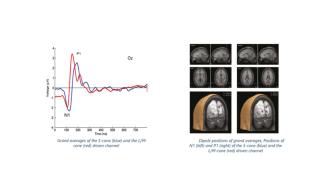
The retinal stages of color processing have been extensively researched in recent years. Of particular interest is the link between color perception disorders and various pathologies of the visual sense. Using the example of glaucoma, a diagnostic relevance could already be shown in own work. In contrast, the cortical stages, especially the spatial distribution of visual processing are less understood. It is known that many cells in the visual cortex respond to color. However, what kind of computations these cells perform on their visual input, and what are their temporal and spatial properties are still under debate. In this study, we aimed to analyze the neuronal sources of visually evoked potentials using selective color opponent channel stimulations. The study was conducted on 10 healthy subjects (6 male, 4 female, 1 eye each, mean age 25.5±5.1 yr). All subjects were free of eye diseases. Normal color vision was checked with Ishihara and Stilling-Velhagen plates. To modulate selective activity in the S-cone and L/M-cone driven channels (the related cells are: short-wavelength absorbing: S-cone, medium-wavelength absorbing: M-cone, long-wavelength-absorbing: L-cone) we presented two silent substitution flash sequences (total of 200 stimuli) in a full circle with a diameter of 22°. Electroencephalography was performed using a 64-channel amplifier system (Theraprax, neuroConn GmbH, Germany) in combination with Ag/AgCl ring electrodes (B10-S-200, EASYCAP GmbH, Germany). The preprocessed data were analyzed with source localization software (CURRY v4.6, Compumedics, USA). For each subject a realistically shaped volume conductor was constructed by a boundary element model. Our findings showed that neural processing occurs in the same areas of the visual cortex for stimuli with different spectral properties. The signals of S- and L/M-cone driven channels are transmitted in distinct pathways to the cortex. Thus, the observed latency differences might be caused by different anatomical and functional properties of these pathways (see graphic).
19.10.2021
Conference paper – 55th Annual Conference of the German Society for Biomedical Engineering (BMT 2021)
“Dry EEG recordings for detection of left, right hand, tongue and feet movements”
Milana Komosar¹, Uwe Graichen1,2, Patrique Fiedler1, Jens Haueisen1
1Biomedical Engineering, TU Ilmenau, Ilmenau, Germany
2Karl Landsteiner University of Health Sciences, Krems an der Donau, Austria
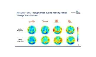
Brain-computer interfaces (BCI) are powerful tools that can be particularly helpful for people with motor disabilities. Recently developed dry EEG sensors could significantly simplify the use of EEG and greatly expand its field of application. In the presented study, EEG data recorded during real and imaginary movements of the left and right hand, tongue and both feet were analysed. The study demonstrated that with the help of EEG systems equipped with dry sensors, BCI technologies can be applied outside of laboratories, non-invasively, quickly, and easily.


